5 Things You Should Know About Persistent Fetal Vasculature
Persistent fetal vasculature was first described as persistent hyperplastic primary vitreous in 1955. The name was later changed to the more medically descriptive persistent fetal vasculature (PFV) in 1997. PFV occurs when the stalk (fetal vessels) that usually regresses during eye development in utero does not retract like it should, if at all. In PFV, light is unable to reach the retina due to the stalk blocking the way. This is the cause for poor or no vision. PFV commonly occurs with microphthalmia and congenital cataracts.
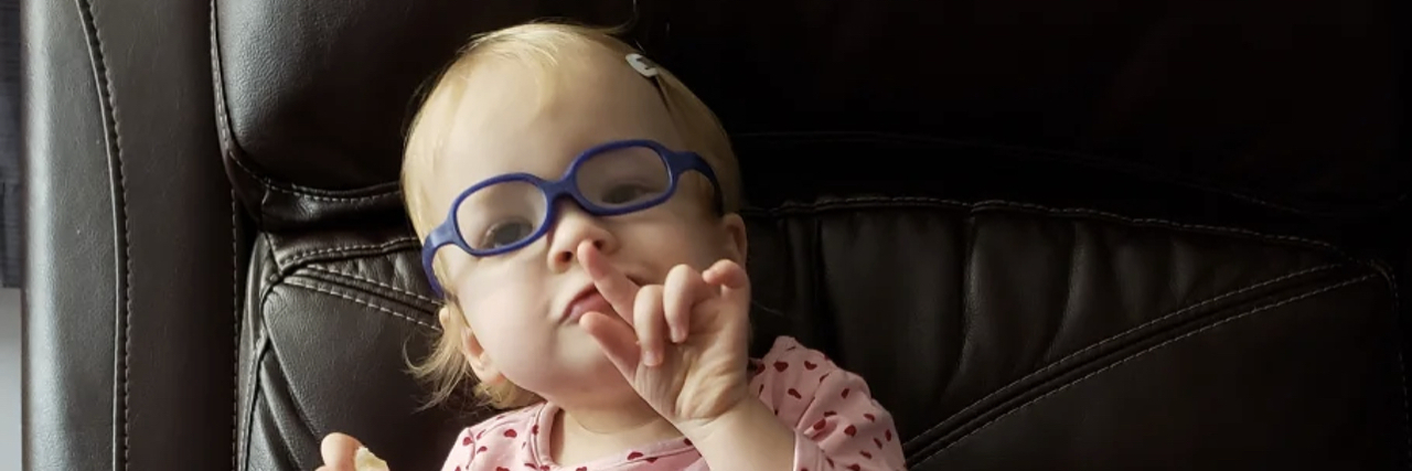
Scarlett got her diagnosis at 6 days old after not opening her right eye at all since her birth. They quickly ruled out retinoblastoma and gave her a diagnosis of microphthalmia, congenital cataract, and PFV. Using ultrasound, it was determined that her stalk never regressed at all. She was completely blind in her right eye and would require a prosthetic eye due to her other conditions.
The five things you should know about PFV.
1. Diagnosis almost always occurs within the first few years of life.
PFV is typically diagnosed as an infant or toddler. If the condition is not diagnosed at birth, other symptoms such as a lazy eye can lead to a true diagnosis of persistent fetal vasculature. This condition is congenital, meaning you are born with it.
2. Symptoms vary depending on severity.
Nystagmus (abnormal eye movements), strabismus (crossed eyes), and amblyopia (“lazy eye”) are common symptoms but each case is different.
Our case was diagnosed after an ultrasound to check the structure of Scarlett’s eye because it is abnormally small (microphthalmia).

3. Misdiagnosis and delayed diagnosis are common.
Parents often notice something is off from birth but are told it is just part of birth trauma or it takes time for things to appear normal. Ideally, diagnosis happens after an eye exam. This exam would warrant a trip to an ophthalmologist where they will request an ultrasound, CT or MRI to confirm their diagnosis of PFV. Exams under anesthesia are also common when the patient is an infant/toddler to check on the eye structure and healing (if surgery was performed).
I know parents who had their concerns dismissed, and went directly to a pediatric ophthalmologist. Other parents would get a diagnosis of a lazy eye only to discover as time went on that PFV was the underlying cause of their child’s symptoms.
4. What treatments exist for the condition?
Each case is individual so treatments will vary. If the patient is a good candidate for vitreoretinal surgery then it is recommended to be done as early as possible. Approximately half of surgical cases gain useful vision in the PFV eye.
After surgery, a patching regimen will be given to help build vision in the affected eye. With patching, an adhesive patch is placed over the stronger eye which in turn makes the weaker eye work harder at gaining vision. This can be challenging to do with children but the reward can be great. Vision can be rehabilitated from just light perception to truly useable vision.
If the case is severe, no treatment or enucleation (removal of the eye) may be recommended. If the eye is removed, a prosthetic eye will be used to maintain the eye orbit structure as well as for cosmetic purposes.
No matter which treatment is done, glasses will be a necessity to protect the eye (or eyes). Often times there will be no prescription in the stronger eye but a prescription for the weaker eye.
5. You are never alone.
Despite only occurring in 1 out of approximately 10,000 births, there is a great online PFV community! From connecting with others through social media to finally meeting in person there are so many opportunities to connect. People with PFV on Instagram and the PHPV Facebook Group have brought together many within the PFV community. In the beginning of your PFV journey, it can feel so lonely. However, I promise you are not. There is a whole PFV family out there and we are pretty awesome.

