
A Beautiful Unpruned Mind-The Neuroscience behind ADHD
Please be patient with ADHD people as they may take longer processing some information, this is also why they deal with overwhelm.
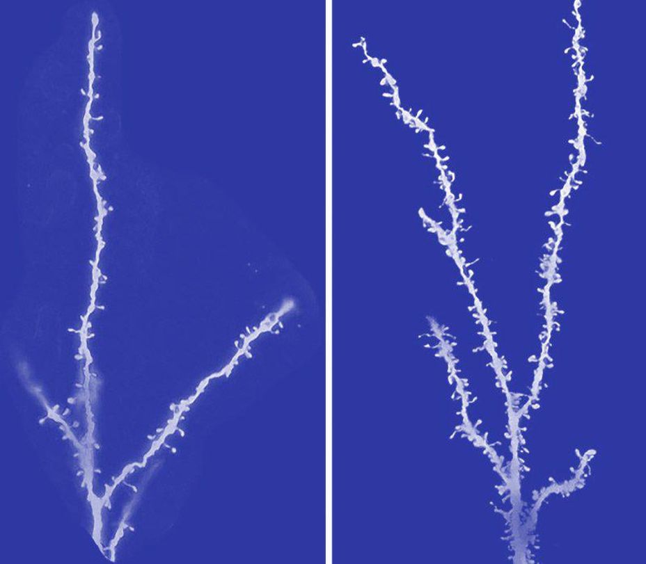
(Image Source Autism -Synaptic Pruning Deficits. On the left you will see a neurotypical synaptic pruning and on the right an ADHD neurodivergent shows less neuron synaptic pruning.)
A Beautiful Unpruned Mind
The human brain is an intricate network of neurons, which communicate through synapses—tiny bridges that allow electrical and chemical signals to pass between cells. During early development, an overabundance of synapses is formed, creating vast neural pathways. However, as we grow, a process known as synaptic pruning takes place. This natural refinement removes unnecessary connections, streamlining brain function and enhancing efficiency (Abitz et al., 2007).
Yet, for some individuals, this pruning process doesn’t occur as extensively as in neurotypical brains. One such group is the neurodiverse or autistic individuals, whose brains retain a higher density of synapses (Tang et al., 2014). While this divergence in neural architecture contributes to characteristic autistic traits, it can also be the foundation for extraordinary cognitive abilities, often linked to savant syndrome.
Synaptic Pruning: The Brain’s Gardener
In infancy and early childhood, the brain experiences a surge in synapse formation, creating a dense web of neural connections. As a child matures, the brain refines itself by eliminating less-used pathways, enhancing efficiency and specialization. Research suggests that by adulthood, neurotypical individuals may have 41% fewer neurons than at birth (Abitz et al., 2007).
However, in autistic brains, this pruning is significantly reduced. Studies indicate that by late childhood, synaptic density decreases by only about 16%, compared to roughly 50% in neurotypical individuals (Tang et al., 2014). This preservation of synapses means that autistic individuals often have heightened connectivity in certain brain regions, which can lead to both challenges and unique cognitive strengths.
The Link Between Autism and Savant Abilities
A fascinating consequence of reduced synaptic pruning is the increased likelihood of savant abilities—exceptional skills in areas such as mathematics, music, or memory. While only about 1% of the general population exhibits savant-like traits, approximately 10% of autistic individuals display such abilities, with some estimates suggesting an even higher prevalence (Treffert, 2009).
One explanation lies in the brain’s hemispheric compensation. Research suggests that damage or developmental differences in the left hemisphere, often observed in autistic individuals, may lead to right hemisphere dominance, which is associated with heightened pattern recognition, spatial awareness, and artistic or mathematical talents (Hughes, 2012). Additionally, brain-derived neurotrophic factor (BDNF) plays a role in neuroplasticity and synaptic growth, potentially fostering enhanced neural connections in those with savant abilities (Schenk et al., 2012).
A Different Kind of Intelligence
While autism presents challenges in areas like social communication, it also offers unique cognitive strengths. A mind that retains more connections may have increased access to vast amounts of raw data, leading to exceptional memory, pattern recognition, and problem-solving abilities (Hughes, 2012). Some researchers speculate that this neural wiring may enable individuals to process information in ways that are unavailable to the neurotypical brain.
The Beauty of an Unpruned Mind
The way our brains develop shapes not only how we think but also who we are. While synaptic pruning streamlines efficiency in most individuals, a brain that holds onto its extra connections may unlock abilities beyond the ordinary. Rather than seeing this as a deficit, it may be more fitting to view it as an alternative form of intelligence—one that fosters unique perspectives, remarkable skills, and in some cases, savant-level genius.
by
Carlita Shaw
#adhd, #neurodivergent #neurodiverse #women #WomenWithADHD #Anxiety #Midlifecrisis #BrainInjury #PTSD #burnout
References
Abitz, M., Damgaard, M., Maier, N., Sakata, M., Aida, T., Kawakami, R., … & Pakkenberg, B. (2007). Excess of neurons in the human newborn mediodorsal thalamus compared with that of the adult. Cerebral Cortex, 17(11), 2573-2580. pubmed.ncbi.nlm.nih.gov/17218480
Hughes, J. R. (2012). A review of Savant Syndrome and its possible relationship to epilepsy. Epilepsy & Behavior, 23(4), 288-294.
Schenk, F., Eckert, P., & Hunziker, J. (2012). Brain-derived neurotrophic factor (BDNF) and its role in neuroplasticity. Frontiers in Neuroscience, 6, 12.
Tang, G., Gudsnuk, K., Kuo, S. H., Cotrina, M. L., Rosoklija, G., Sosunov, A., Sonders, M. S., Kanter, E., Castagna, C., Yamamoto, A., Arancio, O., Peterson, B. S., Champagne, F., Dwork, A. J., Goldman, J., & Yue, Z. (2014). Loss of mTOR-dependent macroautophagy causes autistic-like synaptic pruning deficits. Neuron, 83(5), 1131-1143. doi.org/10.1016/j.neuron.2014.07.040
Treffert, D. A. (2009). The savant syndrome: An extraordinary condition. Philosophical Transactions of the Royal Society B: Biological Sciences, 364(1522), 1351-1357Website- Synaptic growth, synesthesia & savant abilities | Embrace Autism
Additional Notes on the Study
Tang, G., and Sonders, M. S. (2014) contributed to a study that explored the relationship between developmental dendritic pruning, elevated mTORC1 signaling, macroautophagy, and autism spectrum disorder (ASD). The study, conducted at Columbia University Medical Center (CUMC), found that loss of mTOR-dependent macroautophagy leads to autistic-like synaptic pruning deficits, characterized by increased dendritic spine density and reduced developmental spine pruning in layer V pyramidal neurons in postmortem ASD temporal lobe
The study by Tang et al. (2014) was supported by the Simons Foundation, the U.S. Department of Defense (TS110056), the Parkinson’s Disease Foundation, the JPB Foundation, and the National Institutes of Health (NIH K01MH096956, R01MH64168, DP2OD001674, R01NS049442). Additional funding came from the American Heart Association, and brain tissue samples were provided by the Harvard Brain Bank and the Maryland NICHD Brain & Tissue Bank
(You can find this article with an explainer video, in my blog lifepathfrequencies over on WordPress)
Synaptic growth, synesthesia & savant abilities | Embrace Autism


 5 reactions •
2 comments
5 reactions •
2 comments


